


OVERVIEW
Our handcrafted and customized neural electrode arrays can be divided into two parts: the electrode array and the carrier unit.
The electrode array is the interface to the neural tissue and can consist of recording electrodes like
- single core electrodes
- tetrodes (4 core electrodes)
- heptodes (7 core electrodes)
and stimulation sites like
- optical stimulation fiber,
- electrical stimulation electrodes and
- injection cannulas (drug delivery).
The carrier unit provides the interface between the electrode array and the external instrumentation (preamplifiers).
Each carrier unit includes a specific connector (e.g. Omnetics). The same electrode array can be combined with another carrier unit offering you a high flexibility in the configuration of neural probes to meet your specific experimental requirements.
 Figure 1: Thomas Neural Electrode Array
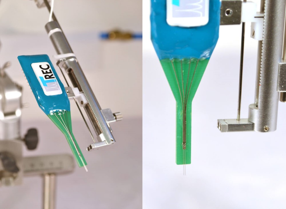 Figure 10: TREC neural probe mounted to a Thomas Motorized Electrode Manipulator (MEM)
Use with Thomas RECORDING Microdrive
Beside the neural probes we also offer bidirectional motorized and software controlled microdrives for moving Thomas neural probes with high precision to different depths of the brain. The Thomas Motorized Electrode Manipulator (MEM) is well suited to drive the Thomas Neural Electrode Arrays with high precision.
Thomas RECORDING Array Electrodes are available in many configurations. Each Array Electrode can be customized for optogenetical applications, drug delivery or electrical stimulation.
Standard Probes
Thomas RECORDING standard Neural Electrode Arrays are manufactured using the quartz glass insulated platinum/tungsten microelectrode technology originally developed at University of Marburg, Germany.
Thomas RECORDING offers a wide variety of Neural Electrode Arrays. The standard Neural Electrode Array consist of a number of single electrodes, tetrodes or heptodes in linear or concentric arrangement.
The arrays can be customized according the users requirements. Standard lateral electrode spacing is 254µm but can be also customized. The protrusion of the electrode fibers from the carrier unit can also be adjusted to the customer´s wishes. So please feel free to select your probe array design.
 Figure 2: TREC Neural Electrode Array with angular electrode arrangement. This picture shows a 5 tetrode array with 20 recording channels.
 Figure 3: TREC Neural Electrode Array with 5 x Tetrode (4 channel electrode fiber). The picture on the left side shows the tetrode tip and a cross section of the tip. The tetrode has a center core and three wires arranged concentrically around the center core. These Neural Electrode arrays are available with single core electrodes, tetrodes (4 metal cores) and heptodes (7 metal cores).
If one is using tetrodes as recording electrodes in a TREC neural array electrode we offer a new designed tetrode spike sorter for convenient data analysis.
Optogenetic Probes
Any of our Neural Electrode Arrays can be configured as an optoelectrode to enable concurrent optical stimulation and neural recording.
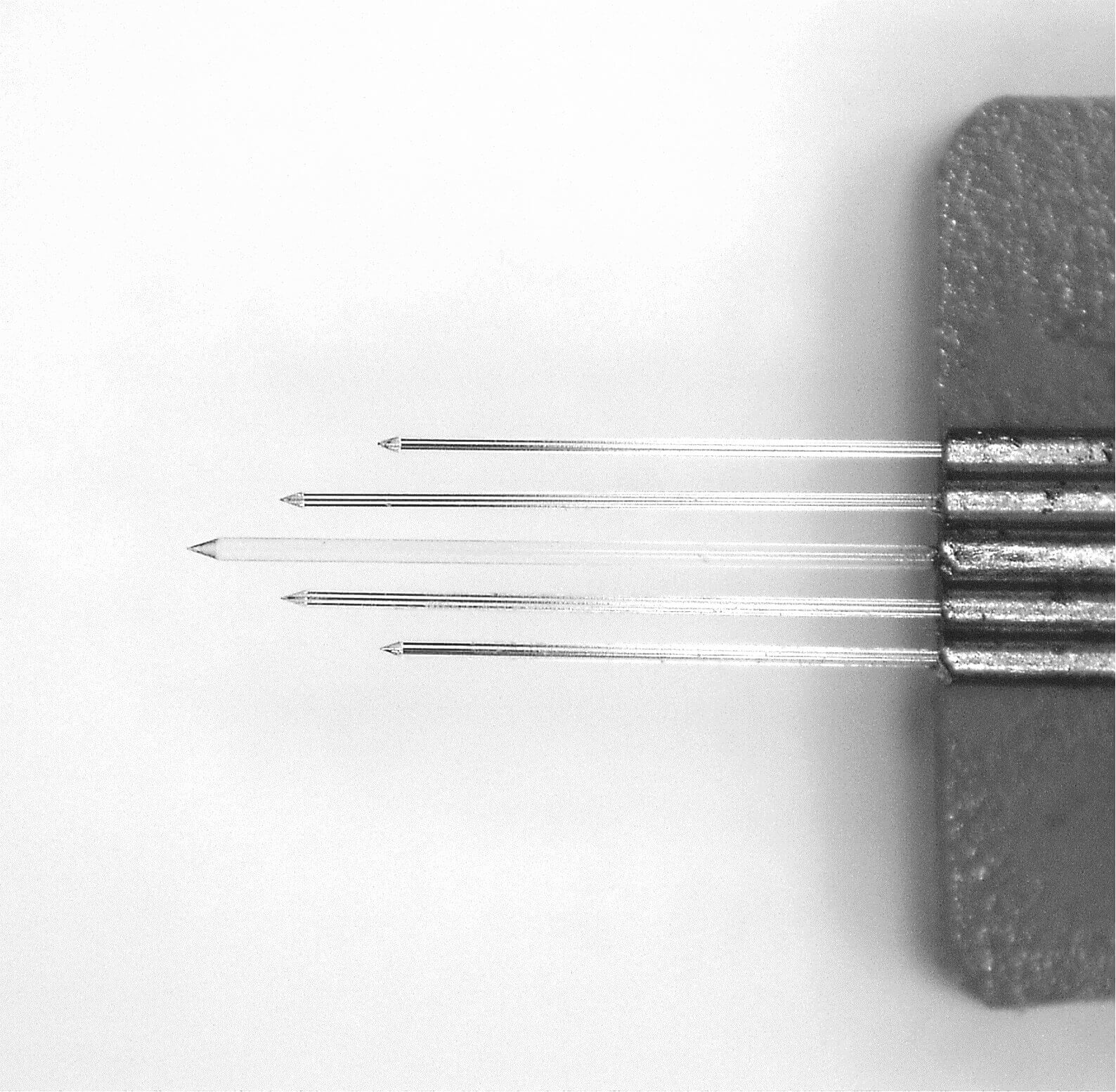 Figure 4: V-Probe The V-Probe arrangement shows 5 fibers (4 tetrodes and one optical fiber in the middle position) with a lateral distance of 254µm.
 Figure 5: Offset Probe
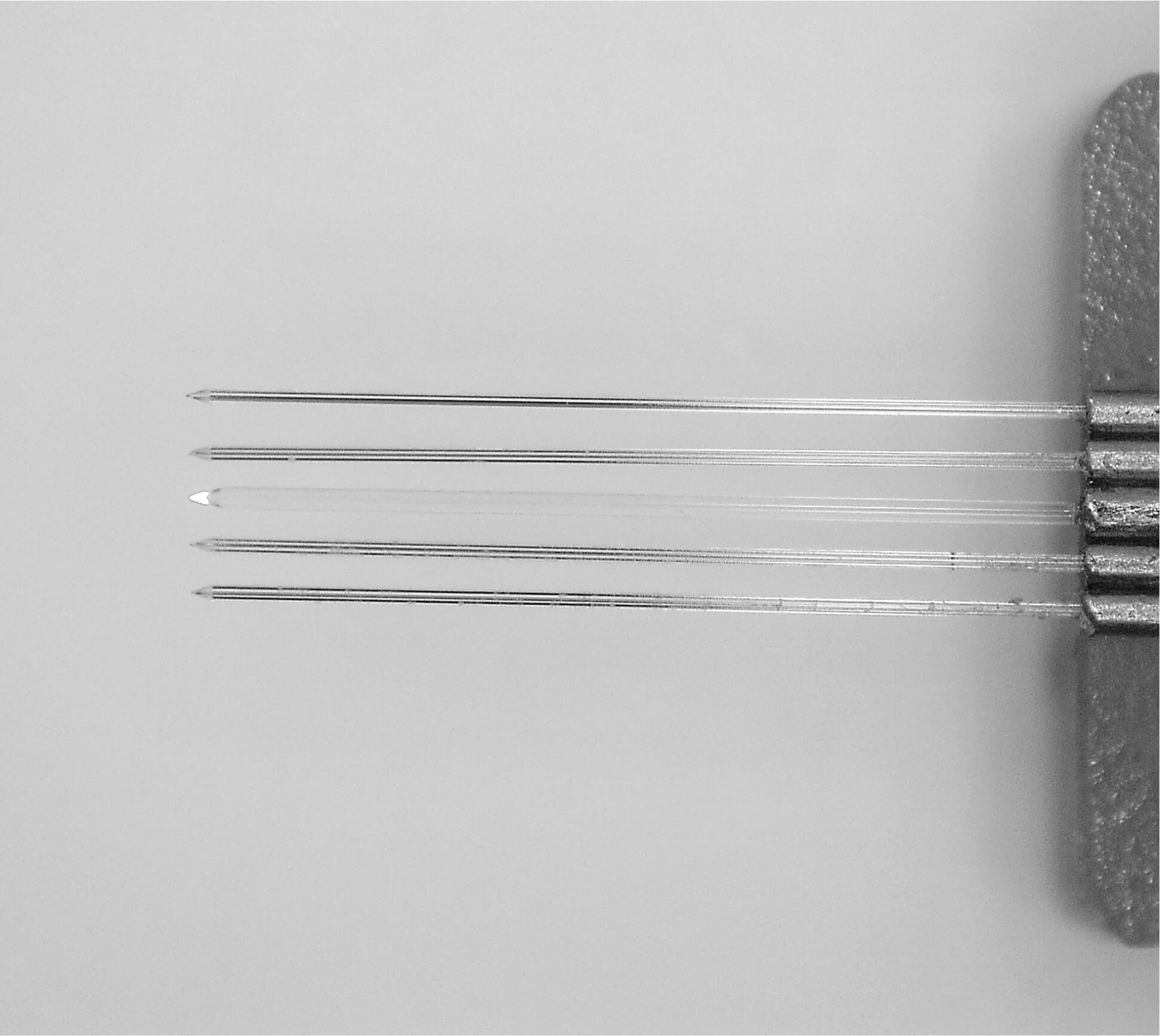 Figure 6: Linear Probe
 Figure 7: Neural Electrode Array with 4 tetrodes (16 recording channels) and one optical fiber for optical stimulation
Drug Delivery Probes
The Thomas Neural Electrode Arrays are available with additional drug delivery channel. By using a Thomas Microinjection System (MIS) precise computer controlled drug injections are possible. This probe combines precise controlled drug injection with extracellular recordings. The fluid interface is compatible with most injection pump systems.
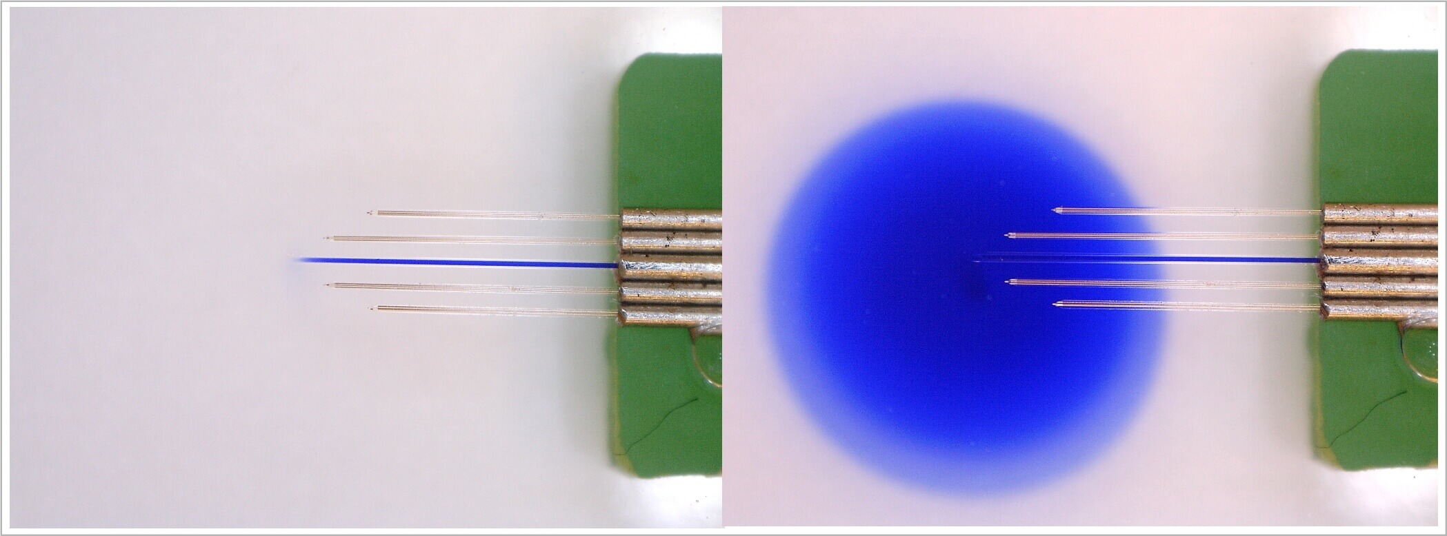 Figure 8: Neural Electrode Array with 4 tetrodes (16 recording channels) and one injection cannula for drug injection
Electrical Stimulation Probes
The Thomas Neural Electrode Arrays are available with additional stimulation electrode. The TREC stimulation electrodes have small outer shaft diameters and low impedance values as well as high charge transfer capacities.

Figure 9: Neural Electrode Array with 4 tetrodes (16 recording channels) and one microstimulation electrode with iridium oxide coating at the tip.
The arrangement of recording electrodes and stimulation sites is customized. So please do not hesitate to contact us for your individual probe design.
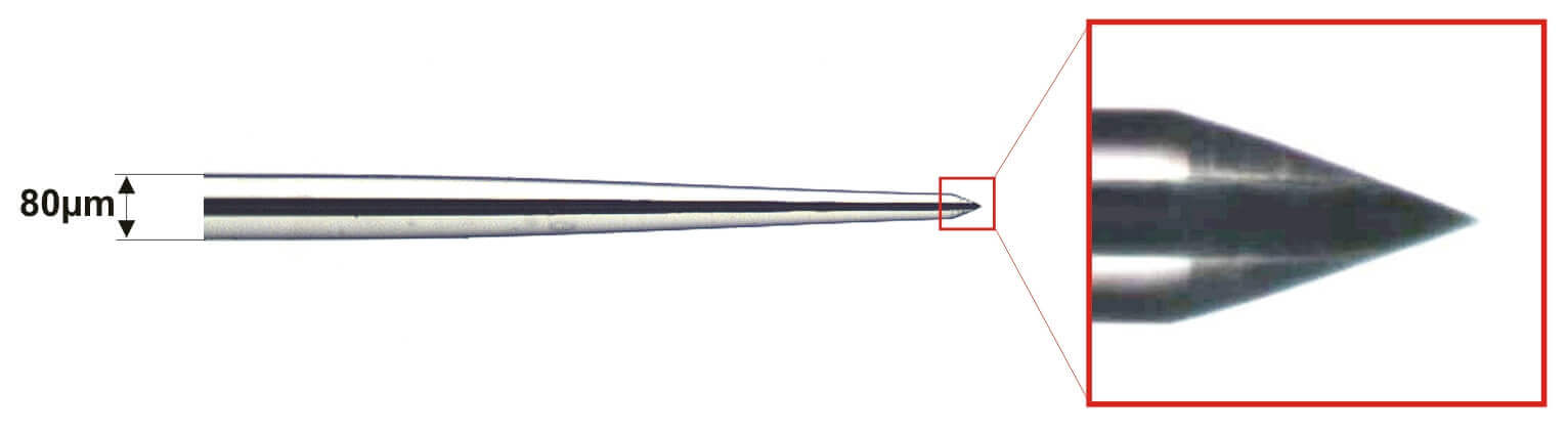 Figure 11: The passage between the glass insulation and the metal part of the electrode tip is so smooth, that only minimal tissue damage is caused when the electrode is introduced in brain tissue.
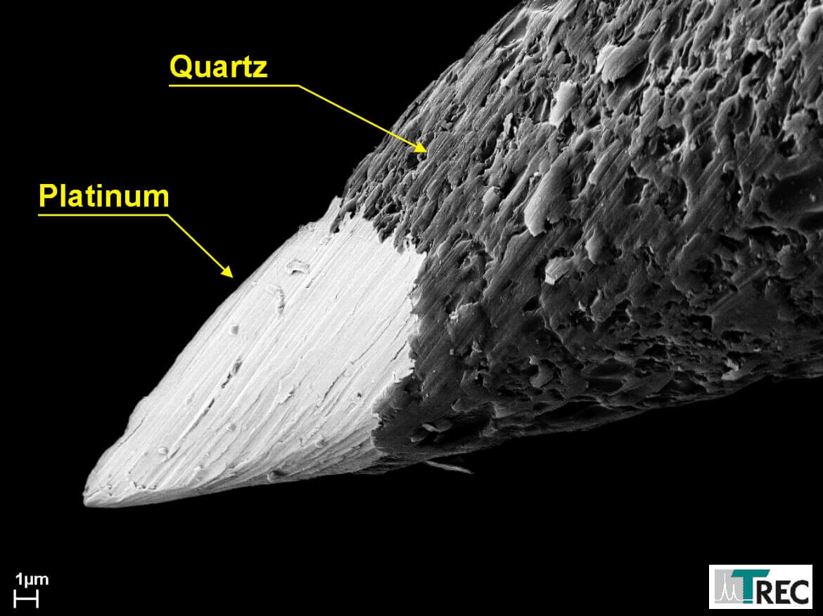 Figure 12: Microgrooves in the metal increase the effective metal area which causes a higher tip-tissue capacity, one reason for the excellent signal to noise ratio of these electrodes.
 Figure 13: Shaft diameter of Thomas fiber electrodes is small and constant over the complete length of the electrode fiber.
 Figure 14: These pictures show a penetration test with Thomas fiber microelectrodes. The electrodes moves straight through the brain over a distance of 40.000µm although the fibers just had 80µm outer diameter. The drawn connecting lines in the rigth photo show that the exit points of the elelctrodes are exactly at the opposite side of the entrance points.

NEWS

PRODUCTS

SOLUTIONS

DISTRIBUTORS
 Sign Up