Tetrodes
(Four Core Fiber Microelectrodes)
The Thomas RECORDING multicore microelectrodes were designed by Heribert Reitboeck and Uwe Thomas in 1987 in the Reitboeck lab at the University of Marburg, Germany. The Thomas tetrode is superior to a single microelectrode due to single unit isolation out of a multi-unit recording. Thomas Tetrodes offer greater spatial resolution, better spike sorting, and improved signal quality compared to single electrodes. This makes them especially useful in complex brain areas where multiple neurons are active in close proximity and need to be recorded simultaneously for accurate analysis. Single electrodes may suffice for simpler recordings but have limitations when it comes to resolving activity from closely spaced neurons or studying larger networks of neurons. Thomas RECORDING tetrodes are ideal for high-precision, multi-unit extracellular recordings, and they allow researchers to gain more insights into neural population dynamics and the functional organization of the brain.
Product Description
Key Features
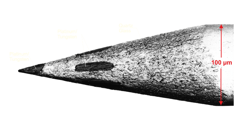
Material: Quartz glass insulated Platinum/Tungsten (95%/5%)
4 metal cores
Fiber diameter of 100µm
Unique material combination
Biocompatible materials
Well suited for acute and also for long term chronic recordings
Very close electrode spacings are possible (down to 100µm) when using Thomas microdrives
very thin shafts minimize tissue damage
Suitable for cortical as well as deep brain recordings
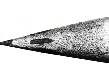
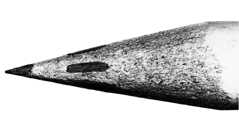
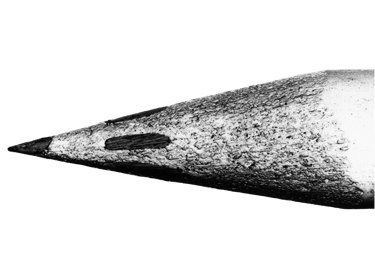
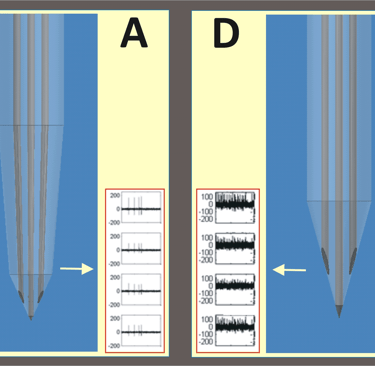
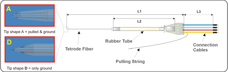
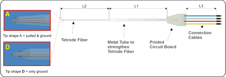
Tetrode Tip Shapes
Thomas RECORDING tetrodes are available with two different tip shapes A and D (see picture on the right side). Tip A (pulled & gound tip) has recording contacts that are closer together and have smaller contact areas, resulting in higher impedance values per contact. This means that each tetrode recording contact has a small recording sphere. Therefore electrode tip is very well suited for signal recordings in brain regions with higher cell density. It allows for better isolation of individual units from a multi-unit recording. In contrast, a Thomas RECORDING tetrode with tip shape D (only ground) is very well suited for brain regions with normal cell density. Thomas RECORDING tetrodes are the only tetrodes on the market whose recording properties can be adapted to different cell densities in different regions of the brain!

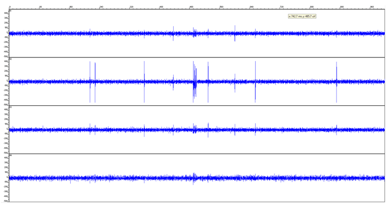
Recording Quality
The picture on the right side shows an extracellular recording with a Thomas RECORDING tetrode from rat brain.


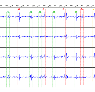
The picture on the left side shows an extracellular recording with a Thomas RECORDING tetrode in subthalamic nucleus of an anesthetized dystonia patient, University Hospital Heidelberg. One can discern up to three different units out of the multi unit tetrode recording (unit A, B and C) based on the stereotrode effect.
Ordering Information
Thomas RECORDING tetrodes are available for Thomas microdrives with rubber tube drive (e.g. Mini Matrix and Eckhorn Matrix ) or for motorized and manual single shaft electrode microdrives (e.g. Thomas MEM).
Tetrodes for Thomas Rubber Tube Microdrives
(Mini Matrix, Eckhorn Matrix, Thomas Matrix)
Tetrodes for other Microdrives & Manipulators
Article numbers:
AN000252 (Eckhorn Matrix, Tip shape D)
AN000253 (Eckhorn Matrix, Tip shape A)
AN000258 (Mini Matrix, Tip shape D)
AN000259 (Mini Matrix, Tip shape A)
Article numbers:
AN000020 (Tip shape D)
AN000021 (Tip shape A)
AN000402 (Tip shape D, with ground wire connected to metal tube)
Selected Publications
[1] Kaneko H, Suzuki SS, Okada J, Akamatsu M. Classifying neuronal spikes from multiunit recording by using a multisite electrode.18th Annual International Conference of the IEEE. 1996. pp. 1500-1501.
[2] Tamura H, Kaneko H, Fujita I. Quantitative analysis of functional clustering of neurons in the macaque inferior temporal cortex. Neuroscience Research 2005; 52: 311-322.
[3] Tamura H, Kaneko H, Kawasaki K., Fujita I. Presumed Inhibitory Neurons in the Macaque Inferior Temporal Cortex: Visual Response Properties and Functional Interactions with Adjacent Neurons. J Neurophysiol 2004; 91: 2782-2796.
[4] Schmidt EM. Electrodes for many single neuron recordings. In: Nicolelis MAL (ed).Methods for Neural Ensemble Recordings. New York: CRC Press LLC; 1999. pp. 1-23.
[5] Reitboeck HJ. Fiber microelectrodes for electrophysiological recordings. J Neurosci Methods 1983; 8: 249-262.
[6] Eckhorn R, Thomas U. Fasermikroelektroden für technische und medizinische Anwendungen. Hannover Messe. 1987.
[7] Swadlow HA, Bereshpolova Y, Bezdudnaya T, Cano M, Stoelzel CR. A multi-channel, implantable microdrive system for use with sharp, ultra-fine "Reitboeck" microelectrodes. J Neurophysiol 2005; 93: 2959-2965.
[8] Tammer R, Ehrenreich L, Boretius S, Watanabe T, Frahm J, Michaelis T. Compatibility of glass-guided recording microelectrodes in the brain stem of squirrel monkeys with high-resolution 3D MRI. J Neurosci Methods 2006; 153: 221-229.
[9] Adams DL, Horton JC. Monocular Cells Without Ocular Dominance Columns. J Neurophysiol 2006; 96: 2253-2264.
[10] Mountcastle VB, Reitboeck HJ, Poggio GF, Steinmetz MA. Adaptation of the Reitboeck method of multiple microelectrode recording to the neocortex of the waking monkey. J Neurosci Methods 1991; 36: 77-84.
[11] Reitboeck HJ. A 19 channel matrix drive with individually controllable fiber microelectrodes for neurophysiological applications. IEEE Transactions on Systems, Man and Cybernetics 1983; 13: 676-683.
[12] Harris KD, Henze DA, Csicsvari J, Hirase H, Buzsaki G. Accuracy of Tetrode Spike Separation as Determined by Simultaneous Intracellular and Extracellular Measurements. J Neurophysiol 2000; 84: 401-414.
[13] Gray CM, Maldonado PE, Wilson M, McNaughton B. Tetrodes markedly improve the reliability and yield of multiple single-unit isolation from multi-unit recordings in cat striate cortex. J Neurosci Methods 1995; 63: 43-54.
[14] Buzsaki G. Large-scale recording of neuronal ensembles. Nat Neurosci 2004; 7: 446-451.
[15] Rebrik SP, Tzonev S, Miller KD. Analysis of Tetrode Recordings in Cat Visual System.CNS. 2007.
[16] Vollgraf R, Munk M, Obermayer K. Optimal filtering for spike sorting of multi-site electrode recordings. Network 2005; 16: 85-113.
[17] Menne KML. Feature extraction from realistically simulated and recorded multisite neuronal signals. Medical University Luebeck, Germany; 2002.
[18] McNaughton B, Barnes CA, O´Keefe J. The stereotrode: a new technique for simultaneous isolation of several single units in the central nervous system from multiple unit records. J Neurosci Methods 1983; 8: 391-397.
[19] Hoehl D. Mechanical evaluation of quartz lass insulated fiber electrodes. unpublished report; 2003.
[20] Eckhorn R, Thomas U. A new method for the insertion of multiple microprobes into neural and muscular tissue, including fiber electrodes, fine wires, needles and microsensors. J Neurosci Methods 1993; 49: 175-179.
[21] Baker SN, Philbin N, Spinks RL, Wolpert DM, MacManus DG, Pauluis Q et al. Multiple single unit recording in the cortex of monkeys using independently moveable microelectrodes. J Neurosci Methods 1999; 94: 5-17.
[22] Wilms M, Eckhorn R. Spatiotemporal receptive field properties of epiretinally recorded spikes and local electroretinograms in cats. BMC Neuroscience 2005; 6: 1-14.
[23] Henze DA, Borhegyi Z, Csicsvari J, Mamiya A, Harris KD, Buzsaki G. Intracellular Features Predicted by Extracellular Recordings in the Hippocampus In Vivo. J Neurophysiol 2000; 84: 390-400.
[24] Logothetis NK, Kayser Ch, Oeltermann A. In Vivo Measurement of Cortical Impedance Spectrum in Monkeys: Implications for Signal Propagation. Neuron 2007; 55: 809-823.
[25] Emondi AA, Rebrik SP, Kurgansky AV, Miller KD. Tracking neurons recorded from tetrodes across time. J Neurosci Methods 2004; 135: 95-105.
[26] Hutchinson W, Lozano A, Davis K, Saint-Cyr JA, Lang AE, Dostrovsky J. Differential neuronal activity in segments of globus pallidus in Parkinson's disease patients. neuroreport 1994; 5: 1533-1537.
[27] Lozano A, Hutchinson W, Kiss Z, Tasker R, Davis K, Dostrovsky J. Methods for microelectrode-guided posteroventral pallidotomy. J Neurosurgery 1996; 84: 194-202.
[28] Engel AK, Moll CK, Fried I, Ojemann GA. Invasive recordings from the human brain: clinical insights and beyond. Nat Rev Neurosci 2005; 6: 35-47.
[29] Chen ChCh, Kühn AA, Trottenberg T, Kupsch A, Schneider GH, Brown P. Neuronal activity in globus pallidus interna can be synchronized to local field potential activity over 3–12 Hz in patients with dystonia. Experimental Neurology 2006; 202: 480-486.
[30] Kühn AA, Trottenberg T, Kivi A, Kupsch A, Schneider GH, Brown P. The relationship between local field potential and neuronal discharge in the subthalamic nucleus of patients with Parkinson's disease. Experimental Neurology 2005; 194: 212-220.
[31] Trottenberg T, Fogelson N, Kühn AA, Kivi A, Kupsch A, Schneider GH et al. Subthalamic gamma activity in patients with Parkinson's disease. Experimental Neurology 2006; 200: 56-65.
[32] Trottenberg T, Kupsch A, Schneider GH, Brown P, Kühn AA. Frequency-dependent distribution of local field potential activity within the subthalamic nucleus in Parkinson's disease. Experimental Neurology 2007; 205: 287-291.
[33] Hoehl D, Thomas U. TREC SCANNER - Ein neues System zur funktionellen Neuronavigation bei der Therapie neurodegenerativer Erkrankungen. Blackwell Verlag; 2003. p. 13.
[34] Capelle HH, Hutchinson WD, Fromm C, Krauss JK. Mikroelektrodenableitung in der funktionellen Neurochirurgie. In: Krauss JK, Volkmann J (eds).Tiefe Hirnstimulation. Darmstadt: Steinkopff Verlag; 2004. pp. 125-168.
[35] Hirabayashi T, Miyashita Y. Dynamically Modulated Spike Correlation in Monkey Inferior Temporal Cortex Depending on the Feature Configuration within a Whole Object. J Neurosci 2005; 25: 10299-10307.
[36] Soteropoulos DS, Baker SN. Cortico-Cerebellar Coherence During a Precision Grip Task in the Monkey. J Neurophysiol 2006; 95: 1194-1206.
[37] Hargreaves EL, Smith A.C., Brown E.N., Suzuki W.A. Tetrode recordings of learning related neural activity in the primate entorhinal cortex during a location-scene task.Society for Neuroscience 2006. 2006.
[38] Bierer SM, Phillips JO, Fuchs AF, Kaneko ChRS, Ling L, Nie K et al. Neurophysiological Studies of Electrical Stimulation for the Vestibular Nerve. USA: NIH; 2007.
[39] Reich DS. Information Encoding by Individual Neurons and Groups of Neurons in the Primary Visual Cortex. New York: Rockefeller University; 2000.
[40] Emondi AA. Technical Report for Thomas Recording. 2000.
[41] Kaneko H, Tamura H, Suzuki SS. Tracking spike-amplitude changes to improve the quality of multineuronal data analysis. IEEE Trans Biomed Eng 2007; 54: 262-272.
[42] Kaneko H, Tamura H, Kawashima T, Suzuki SS, Fujita I. Efficient Signal Processing of Multineuronal Activities for Neural Interface and Prosthesis. Methods Inf Med 2007; 46: 147-150.
[44] Mechler, F. and Victor, J. D. Dipole characterization of single neurons from their extracellular action potentials. J Comput Neurosci Published online: June 11, 2011[DOI 10.1007/s10827-011-0341-0]. 11-6-2011
[45] Mechler, F., Victor, J. D., Ohiorhenuan, I. E., Schmid, A., and Quin, H. Three-dimensional localization of neurons in cortical tetrode recordings. Journal of Neurophysiology 106[2], 828-848. 2011
Request Information
Location
Winchester Strasse 8
35394 Giessen, GERMANY
Hours
Mo.-Fr. 8:00 a.m - 4:00 p.m.
Central European Time (CET)
Sa. - Su. Closed
info@ThomasRECORDING.com